
2025
Evolus Academy Aesthetics Education on Facial Anatomy
Entrant
Alexander & Turner Inc. Medical Illustration Studio
Category
Advertising - Illustration / Graphic
Client's Name
Evolus Inc.
Country / Region
United States
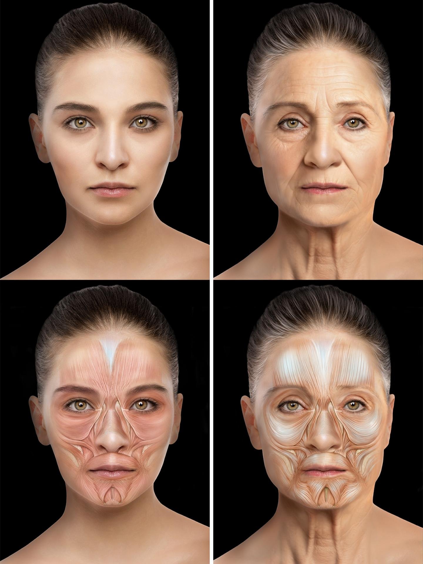
A series of sequential anatomical illustrations demonstrating how facial anatomy changes with age depicting a 25-year old woman aged to 65-years old, developed with the guidance of Professor Sebastian Cotofano M.D., Ph.D, Ph.D. for his educational seminar to teach aging facial anatomy to aestheticians who inject fillers to restore a youthful appearance. Professor Cotofano is recognized globally as the world's leading anatomist in the field of facial aesthetics. The illustrations appear as supporting educational material in a Powerpoint presentation that accompanies a live dissection seminar by Professor Cotofano for Evolus Academy.
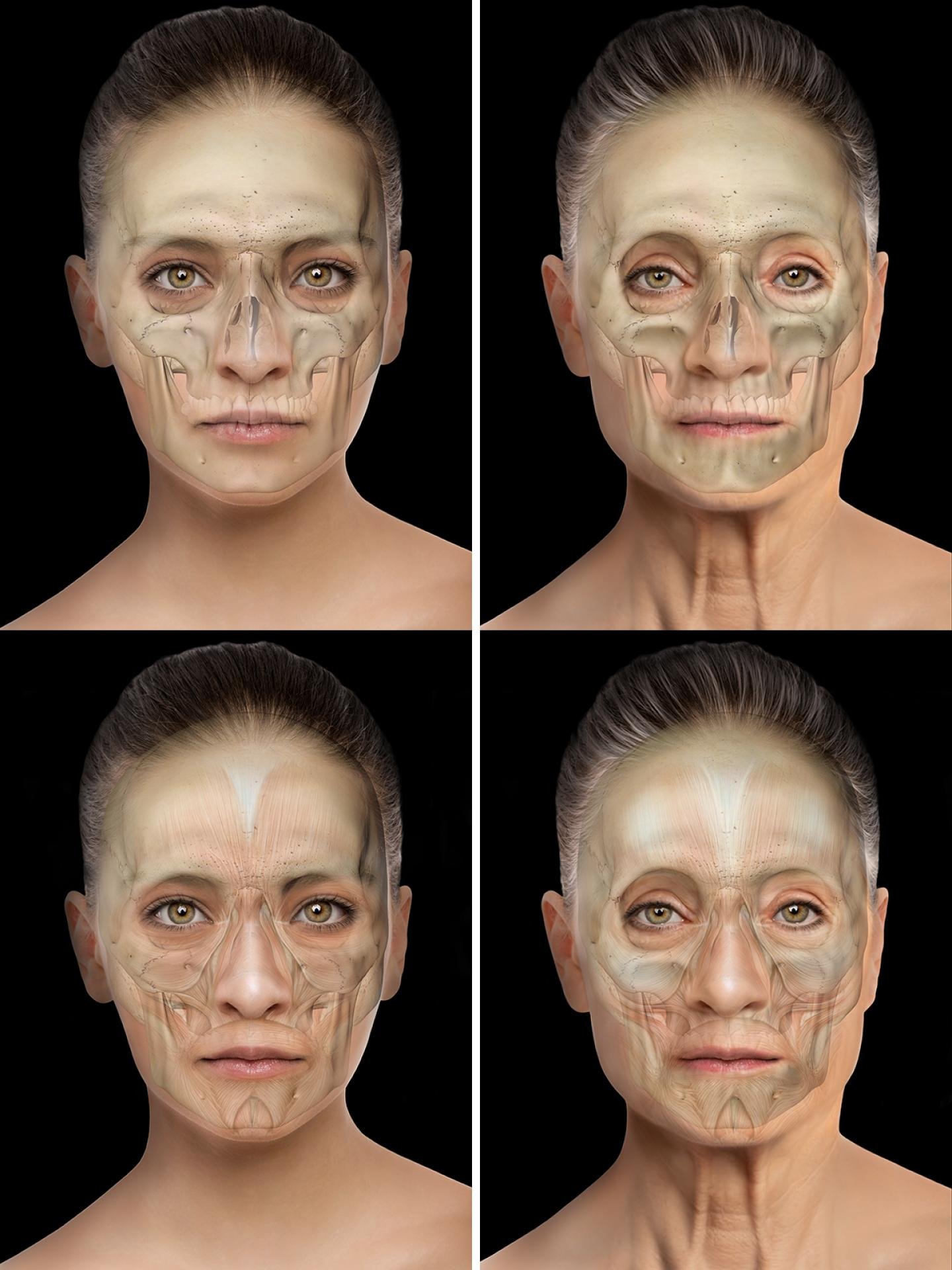
Illustration descriptions: The bone density of the aging skull declines with resorption, making the eye sockets (orbits) larger, wider and longer. The midface, (especially the maxilla) resorbs leading to a reduction in its projection, contributing to the deepening of nasolabial folds on the skin. The mandible becomes more anterior, oblique and shorter.
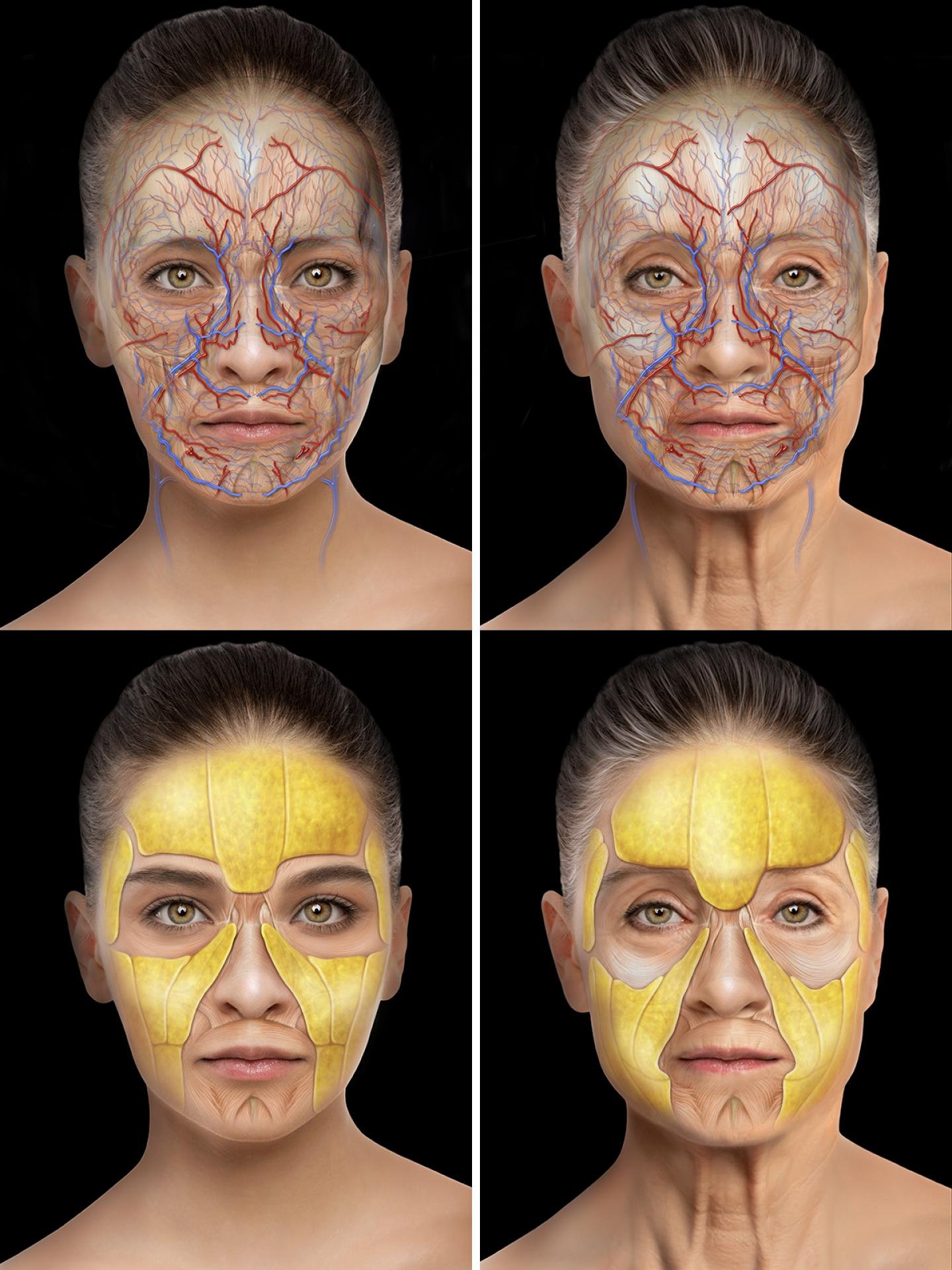
Aging muscles thin, lose elasticity, droop, stretch and become fibrous. Muscle fibers spread out and spaces appear between the muscle fibers, allowing fibrous intrusion. Fat pads sit atop the muscles and travel with the droop of aging muscles. Aging fat pads thin and become slightly smaller, exposing the bony prominences of the skull’s inferior orbital rim, zygomatic arch, and temporal crests.
The skin reflects these deeper changes of the skull, muscles and fat pads. There are increased tear troughs and decreased volume in the temples. Decreased volume in the pyriform fossa causes pronounced nasolabial folds (marionette lines) around the mouth. Jowling develops along the jawline. The loss of collagen and elastin in the skin itself causes wrinkling.
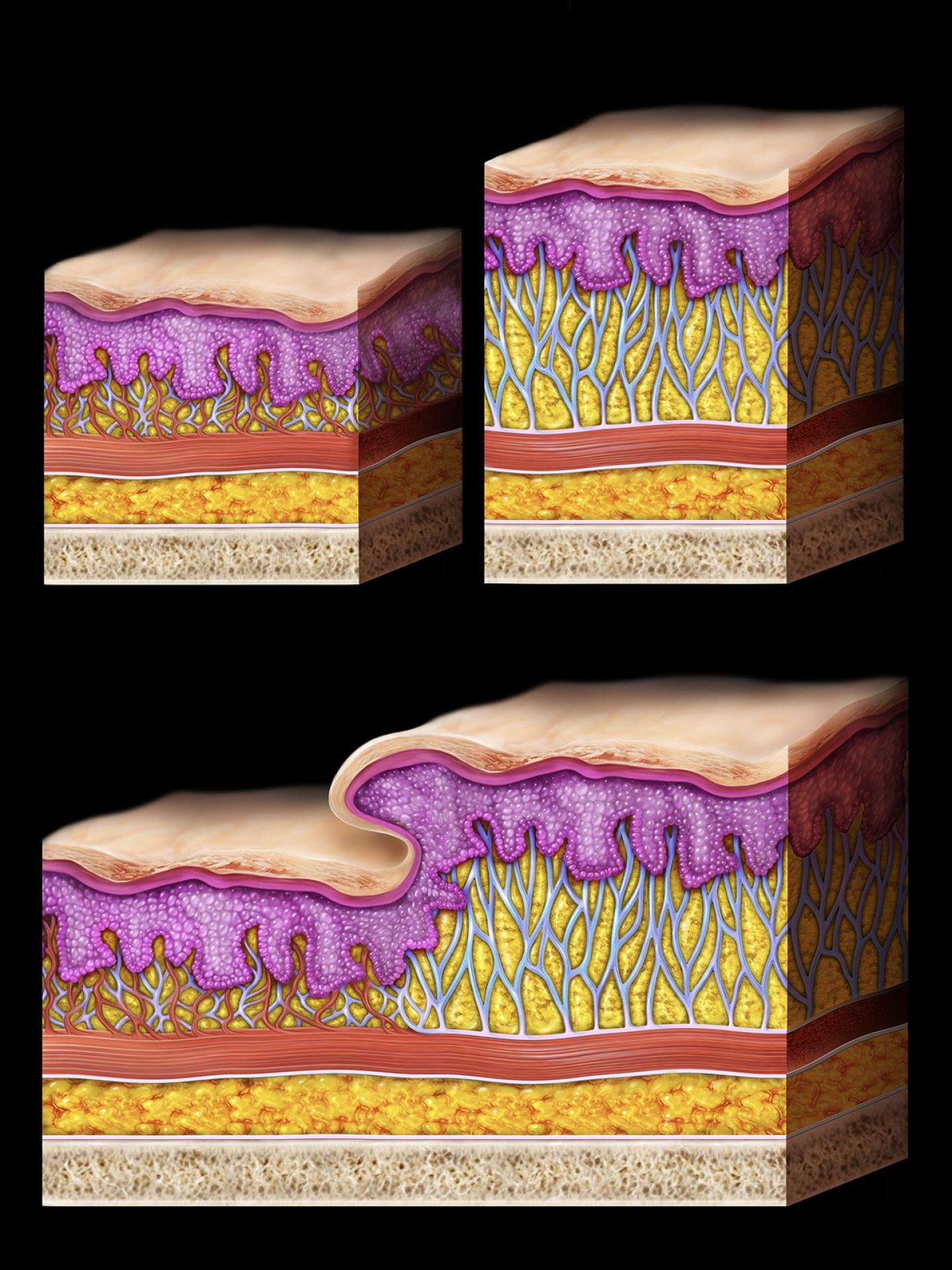
Skin cross sections highlight the difference between the facial medial skin layers and lateral skin layers, crucial to understanding the nasolabial folds. In the facial medial skin layers the retinacula cutis is in the superficial fat layer and is collapsed to closely attach to muscle, allowing for expression and oral movement. In the facial lateral skin layers the retinacula cutis are extended, allowing for skin to act more independently of the muscle. At the nasolabial fold the medial skin layers merges with the lateral skin layers.
Credits

Entrant
R&R Partners
Category
Advertising - New Category
Country / Region
United States

Entrant
American Kidney Fund
Category
Provider & Services - Education
Country / Region
United States

Entrant
Beverly Trading (Shenzhen) Co., Ltd.
Category
Provider & Services - Healthcare
Country / Region
China

Entrant
Zeyu Liu
Category
Interior Design - New Category
Country / Region
United States